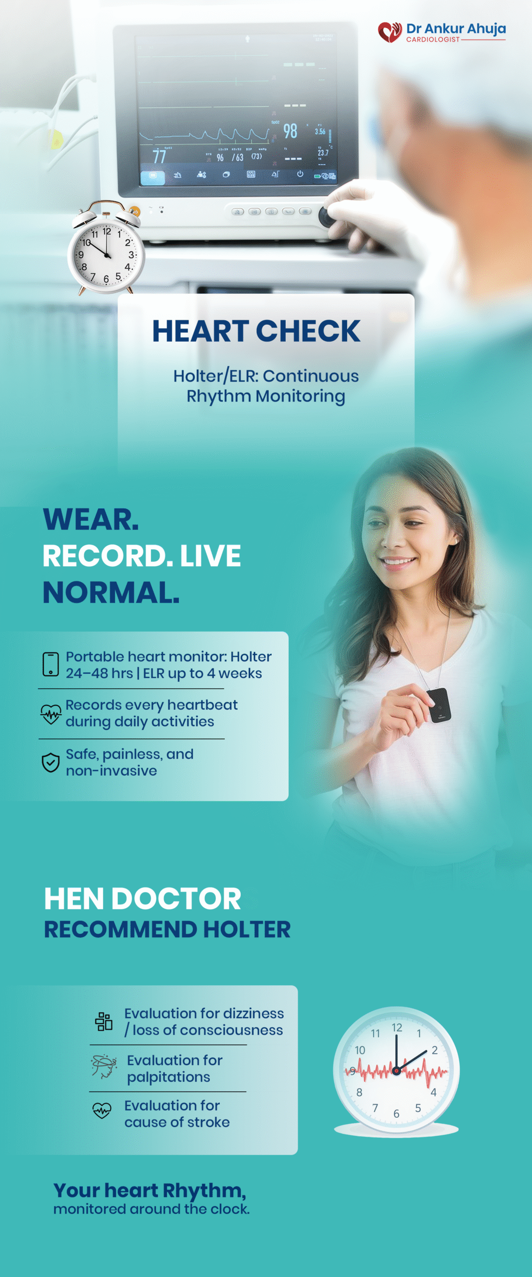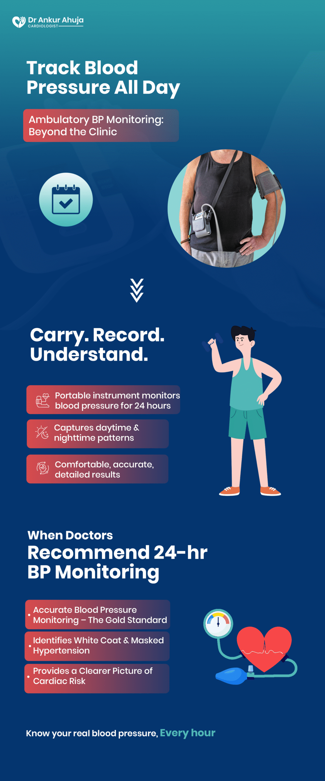
Heart Attack vs Cardiac Arrest: Know the Difference
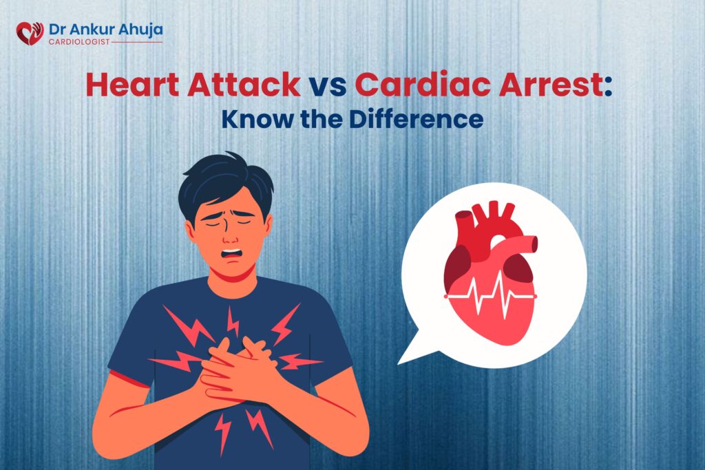
Understanding Heart Attack vs Cardiac Arrest A lot of people use the terms “heart attack” and “cardiac arrest” interchangeably, but […]
Understanding Heart Attack vs Cardiac Arrest A lot of people use the terms “heart attack” and “cardiac arrest” interchangeably, but […]
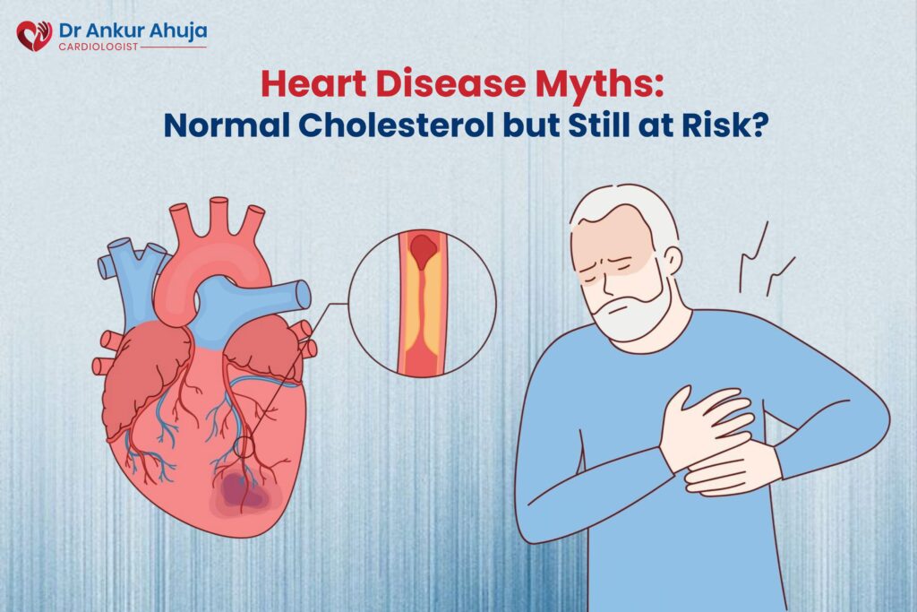
For many individuals, a normal cholesterol report brings relief. It feels like confirmation that the heart is healthy and the
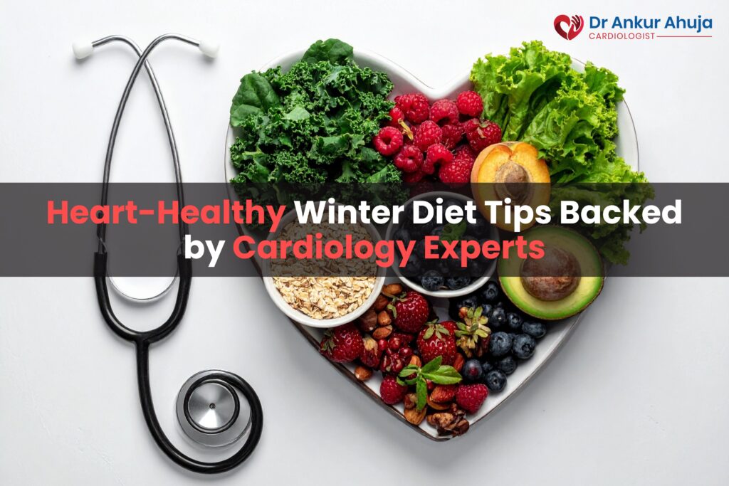
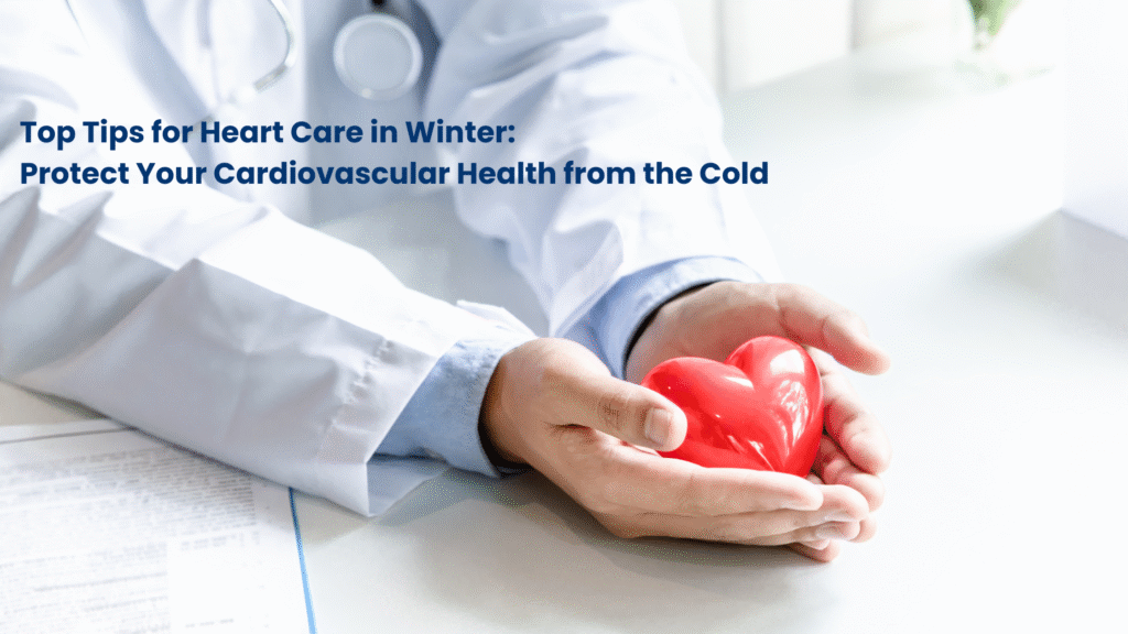
Winter brings comfort foods, reduced physical activity and lifestyle changes that can quietly increase strain on the heart. Cold temperatures
Winter can quietly place extra stress on the heart. Cold temperatures, reduced physical activity, dietary changes, and dehydration all combine
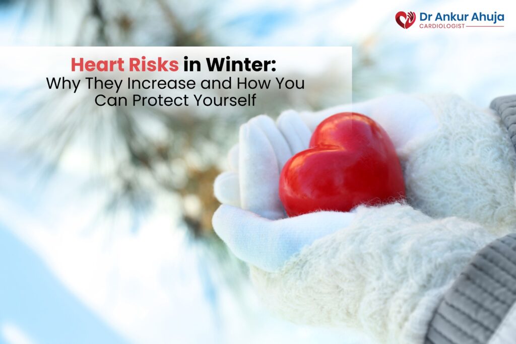
As temperatures drop, many people focus on seasonal flu and infections, but one silent threat often goes unnoticed—Heart Risks in

Cold weather heart health is a serious concern, especially for people with existing cardiovascular conditions. Winter places extra stress on
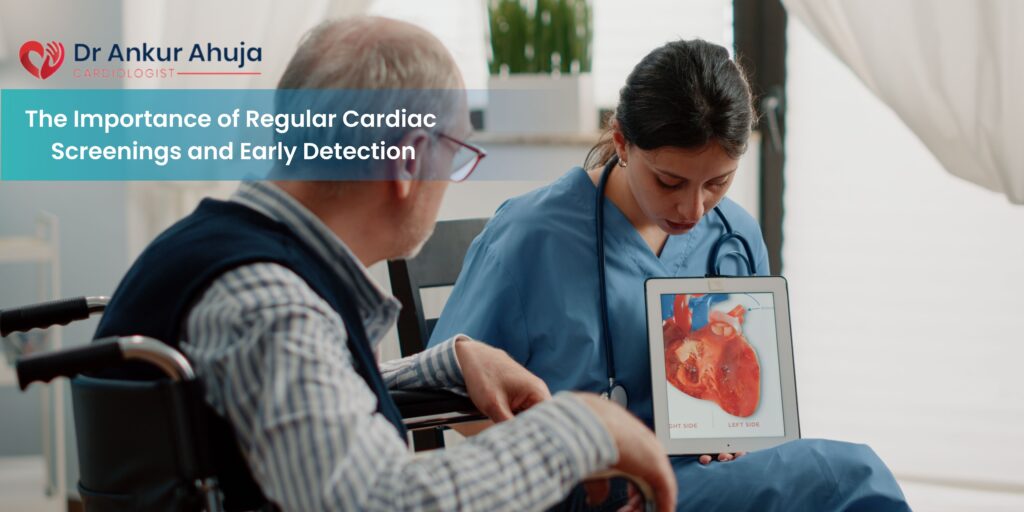
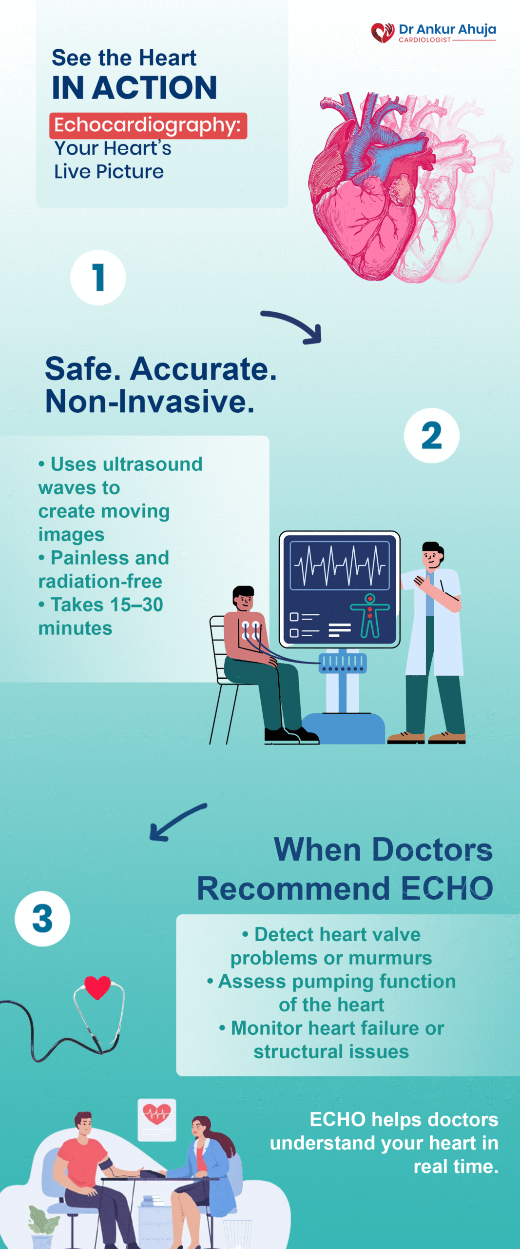
Heart disease remains one of the leading causes of preventable illness and death globally. Despite growing awareness, millions of people
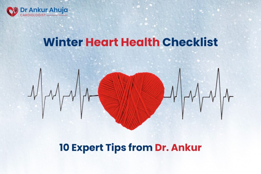
Winter brings cold winds, cozy sweaters, and warm drinks—but it also brings increased risks for heart patients. When it comes

COVID-19 is a rapidly evolving pandemic. Very little is known about it, and most of the information has accumulated from
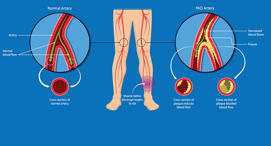
Peripheral arterial disease (PAD) i.e. disease of arteries of the peripheries, is often an unrecognized manifestation of atherosclerosis and other